IASR 43(3), 2022【THE TOPIC OF THIS MONTH】 Toxoplasmosis
PDF download(PDF:578KB)
The topic of This Month Vol.43 No.3(No. 505)![]()
Toxoplasmosis
(IASR Vol. 43 p49-50:March 2022)
Toxoplasmosis is an infectious disease caused by a parasitic protist, Toxoplasma gondii. The prevalence in humans varies by country and age, but it is estimated that at least 1/3 of the human population in the world is infected with T. gondii. Prevalence is particularly high in Brazil, France, and Indonesia. The prevalence in Japan is unknown, but is thought to be around 5-10% (see pp. 51 & 52 of this issue). The disease is not a notifiable disease under the National Epidemiological Surveillance of Infectious Diseases (NESID) program based on the Infectious Diseases Control Law. However, for domestic animals, T. gondii is an important pathogen and is designated as a notifiable infectious disease (target animals: sheep, goats, pigs, and boars) under the Domestic Animal Infectious Disease Prevention Law (see p. 53 of this issue).
Pathogen
T. gondii is an oval to crescent-shaped unicellular organism 2-3 µm wide and 5-7 µm long (Fig. 1). It is a commensal intracellular parasite and can only multiply within the host cell. The life cycle of T. gondii (Fig. 2) consists of sexual reproduction in the intestinal tract of the definitive host, the feline family (Fig. 2, right), and asexual reproduction in the intermediate host, which are all warm-blooded vertebrates including humans and domesticated and wild animals (Fig. 2, left).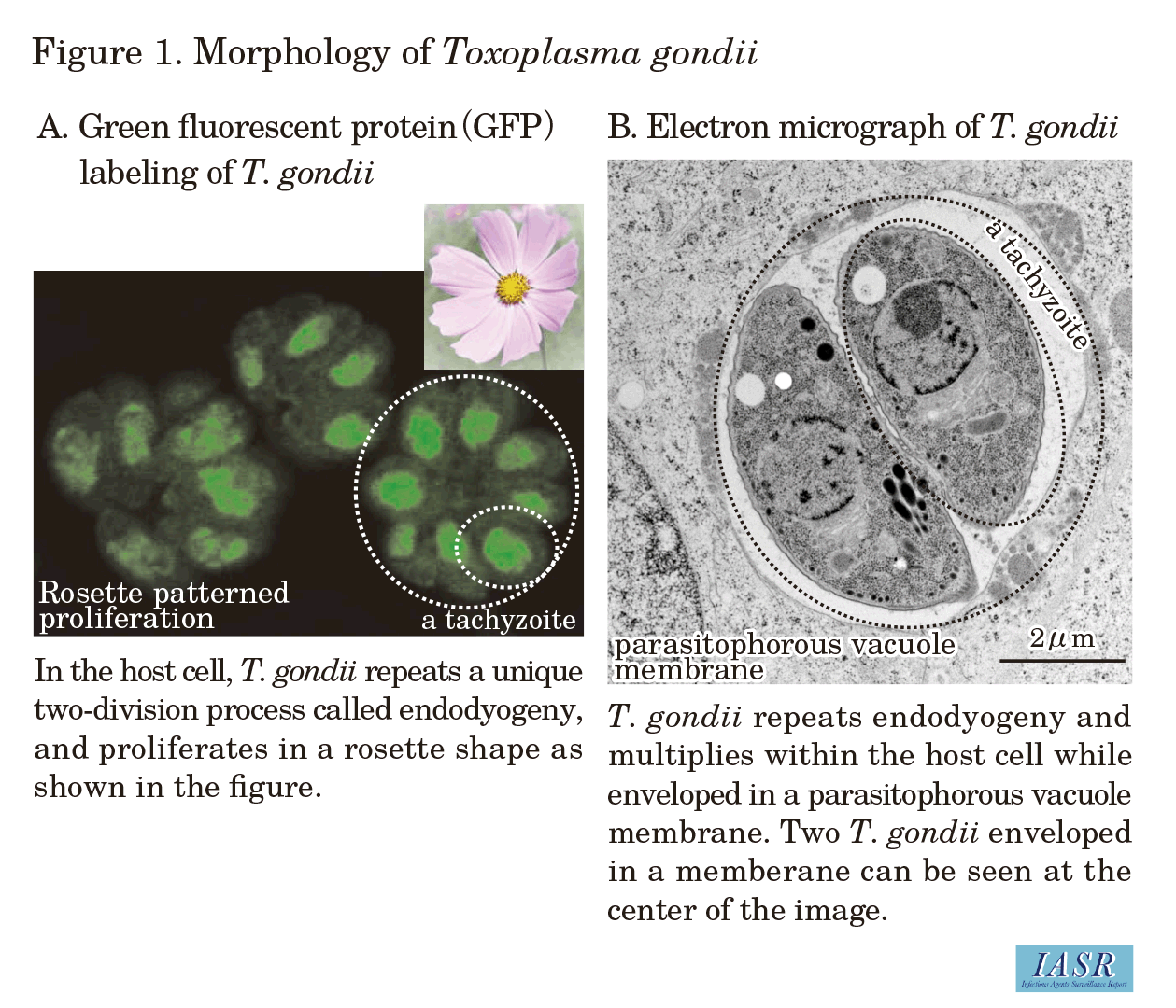
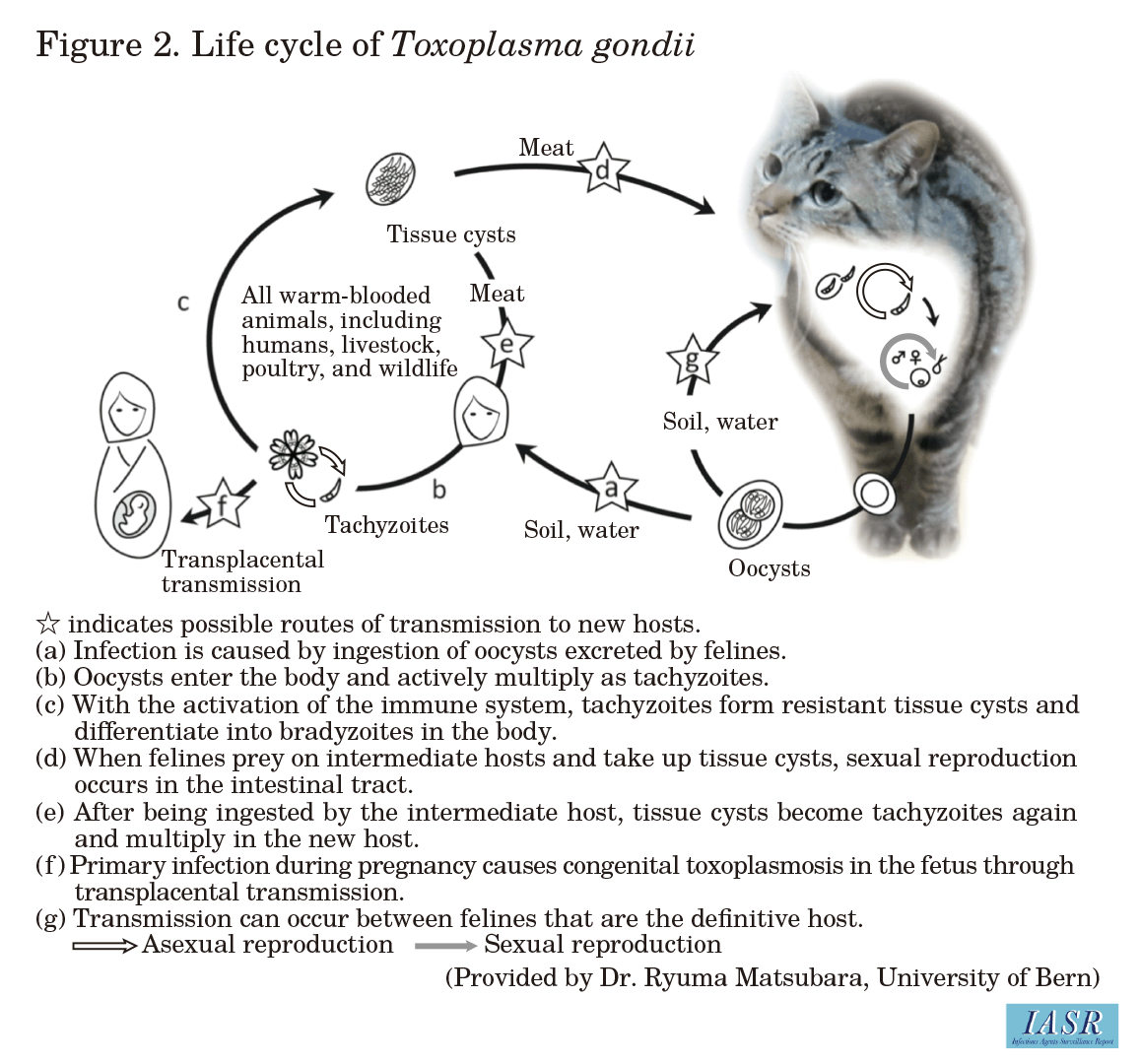
Transmission to intermediate hosts occurs by oral ingestion of tissue cysts formed in other intermediate hosts (Fig. 2e) or oocysts (Fig. 2 a) that have been shed with feces from the definitive host, the feline. The ingested T. gondii enters the intermediate host through the gastrointestinal wall and multiplies actively (Fig. 2 b). If the host is pregnant during the acute infection period, T. gondii may cross the placenta and be transferred to the fetus (Fig. 2 f). Once the host's immune response is initiated, T. gondii forms a solid structure covered with a stable wall (cyst wall) called a tissue cyst (Fig. 2 c), mainly in the central nervous system or muscles, sustaining infection. Growth within the tissue cysts is very slow, and it is diff icult to kill the parasite with antiparasitic drugs (see p. 61 of this issue). T. gondii migrates to a new host and resumes proliferation when an infected intermediate host with tissue cysts is ingested by another intermediate host, such as a human (Fig. 2 e).
When a feline, the definitive host, consumes an infected intermediate host (Fig. 2 d), T. gondii is released from the tissue cysts and differentiates into female and male gametes. The gametes fuse together in the intestinal tract to form immature oocysts that are excreted out of the body with feces. Immature oocysts mature in the external environment. Mature oocysts are highly tolerant to the environment and can survive in soil or water in a stable state for many years (Fig. 2 a).
Route of infection and infection control
As described above, there are two routes of transmission for T. gondii to humans as an intermediate host: oral ingestion of oocysts shed from the feces of the definitive host, the feline, or, of the tissue cysts formed in the central nervous system or muscles of other mammals or birds which act as intermediate hosts. Transmission through the palpebral conjunctiva is also possible, but there is no airborne or transdermal transmission.
In Japan, pork has been conventionally considered to be the main source of oral ingestion of tissue cysts. This is probably because T. gondii infection shows characteristic manifestations of acute signs in pigs, whereas infection in many other domestic animals is mostly asymptomatic, similar to humans. However, since T. gondii can infect not only pigs but all warm-blooded animals, eating raw or undercooked meat of animals, including whale meat or poultry, always carries the risk of infection (see pp. 53 & 54 of this issue). Since clinical symptoms are not readily recognized in animals other than pigs, it is rather diff icult to distinguish infected from uninfected animals and caution is required. Pregnant women or those who may be pregnant should refrain from eating raw meat, and when cooking meat, it should be thoroughly cooked to the center, and the use of cutting boards should be separated by meat vs. other foods. Recently, the possibility that shellfish may retain the oocysts that have been carried from rivers to the ocean, and that shellfish and the marine mammals that feed on them may be a source of infection, has also been discussed (see p. 56 of this issue).
In addition to meat, many cases of infection from water and soil sources have been acknowledged; notably, with outbreaks having been reported for waterborne transmissions, attention should also be paid to the risk of infection from the environment. T. gondii infection from the environment is caused by oocysts in the feces of the definitive host, the feline. Oocysts remain stable and survive in the external environment for many years; additionally, they are resistant to many disinfectants, including hypochlorous acid and ethanol. Contact with soil, such as during gardening or farming, and ingestion of water from sources such as wells or springs, increase the risk of infection. However, because infected cats shed oocysts only for a few days to about 2 weeks after the initial infection, and as it takes at least 24 hours for the shed oocysts to mature and become infectious, daily disposal of feces (within 24 hours) can reduce the risk of contact with an infectious oocyst. There is no need to get rid of a cat for reasons related to pregnancy.
Clinical Symptoms and Diagnosis
The clinical manifestations of toxoplasmosis largely depend on the time of infection and the health status of the infected person. In addition to clinical symptoms, serodiagnosis and genetic testing are the main diagnostic methods (see p. 60 of this issue).
- Congenital toxoplasmosis
If a pregnant woman is infected with T. gondii for the first time, T. gondii can cross the placenta and vertically infect the fetus (see pp. 57 & 65 of this issue). The likelihood of fetal infection increases toward the latter stages of pregnancy; however, if an in utero infection does occur, earlier the stage of pregnancy, higher the severity. The outcome of in utero infection varies widely from subclinical to miscarriage, and the severity varies also among cases of symptomatic infection. The four major recognized signs of congenital toxoplasmosis are hydrocephalus, visual impairment due to choroiditis, intracranial calcification, and psychomotor dysfunction. In addition, congenital toxoplasmosis has been designated as one of the specified pediatric chronic diseases since 2017 in Japan. If infection is suspected in a pregnant woman, the risk of infection in the fetus should be assessed using the IgG or IgM antibody test or the IgG avidity test for the pregnant woman. If the risk is high, diagnosis of fetal infection should be attempted by PCR detection of the protist gene in the amniotic fluid, but this is not definitive. For postnatal diagnosis, serological examination of the infant should be performed after the disappearance of maternal antibodies.
- Acquired toxoplasmosis
(1) Acute infection: Most cases of acquired T. gondii infection in healthy individuals are asymptomatic, and those who develop illness present nonspecific transient symptoms such as fever, malaise, and lymphadenopathy.
(2) Chronic infection: When the active growth of T. gondii is inhibited by the immune system, the infection shifts to a chronic phase. Toxoplasmosis during the chronic phase of infection is considered asymptomatic, but in recent years, it has been reported that toxoplasmosis may increase the risk of behavioral changes in rodents and the development of schizophrenia and other psychiatric disorders in humans (see p. 62 of this issue).
(3) Ocular toxoplasmosis: Toxoplasmosis occurs sporadically in the eye. Symptoms include impaired vision, eye pain, and photophobia (see p. 59 of this issue).
(4) Opportunistic toxoplasmosis: In immunocompromised individuals, chronically infected parasites reactivate and cause severe symptoms such as encephalitis. Clinical manifestations of toxoplasmic encephalitis include altered mental status, convulsions, and visual impairment. Contrast-enhanced CT or MRI of the head shows ring-enhancing lesions. Toxoplasmic encephalitis is diagnosed by PCR detection of the T. gondii gene in spinal fluid, but the sensitivity is low, and a negative result does not rule out infection. In Japan, toxoplasmic encephalitis is one of the 23 indicator diseases of acquired immunodeficiency syndrome (AIDS), a Category V notifiable infectious disease in NESID, and, among cases of patients diagnosed with AIDS in Japan in 2020, it accounted for 1.4% of the Japanese cases and 6.3% of the foreign national cases [2020 Annual Report on HIV/AIDS Surveillance in Japan, the National AIDS Surveillance Committee, Ministry of Health, Labour and Welfare: https://api-net.jfap.or.jp/status/japan/nenpo.html].
Treatment
Pyrimethamine, sulfadiazine, and horinat, which are used overseas for the treatment of toxoplasmosis, are not approved in Japan as at February 2022, but clinical trials for toxoplasmosis and congenital toxoplasmosis are currently underway. To note, these drugs are available through the Research Group on Chemotherapy of Tropical Diseases (see pp. 57 & 59 of this issue). In addition, spiramycin, which has been used in at least 70 countries for the treatment of congenital toxoplasmosis, became commercially available from September 2018 in Japan. Spiramycin has been reported to reduce the severity of congenital toxoplasmosis by 80% and prevent fetal infection by at least 60%, and is expected to improve the clinical environment for treating congenital toxoplasmosis in Japan.
